A corneal abrasion is a cut or scratch on your cornea. The cornea is the clear, protective window at the front of your eye. It lies directly over the colored part of your eye (called the iris).
Many things can cause a corneal abrasion, such as sand, dust, dirt, wood or metal shavings that get in your eye. The cornea can also be scratched by a fingernail, a tree branch or a contact lens. Rubbing your eyes very hard is another way that an abrasion can occur. Sometimes, if a corneal abrasion hasn't healed properly, it can come back weeks or months after the original injury. In some people, the outer layers of the cornea are weak. These people may get a corneal abrasion for no apparent reason.
The cornea is very sensitive, and a corneal abrasion is usually quite painful. You may feel like you have sand or grit in your eye. You may notice tears or blurred vision, or your eye may look red. You may also notice that light hurts your eye. Some people get a headache when they have a corneal abrasion.
If you think something has gotten into your eye, first try to wash out the eye by splashing clean water into it. Your workplace may have an eye rinse station for this purpose. Sometimes, blinking or pulling the upper eyelid over the lower eyelid may remove a particle from under the eyelid. Avoid rubbing your eye. If you or someone else notices something on the white part of your eye, use a soft tissue or cotton swab to gently lift it out of the eye. Don't try to remove something that is directly over the cornea--this might cause more serious damage. If you can't remove the particle or if there doesn't seem to be anything in your eye, call your doctor.
Your doctor will examine your eye for any damage or particles that may be trapped under your eyelid. A yellow-orange dye may be placed on your eye to help your doctor see the abrasion. Your doctor will probably treat the abrasion with eye drops or ointment. You may need to wear a patch on your eye overnight. Most small abrasions heal within 24 hours, but you may need to return to your doctor for another exam the next day.
If you wear contact lenses, you need to be especially careful with a corneal abrasion because you have a higher risk of infection. If you have any of the symptoms described here, call your doctor.
Corneal Dystrophy
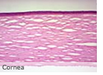
The globe of the eye is made of five layers with the cornea being the transparent front portion. It is also the most sensitive structure in the body due to the density of nerves. The cornea is transparent due to the presence of a regular lattice structure of collagen fibres. Anything which affects this regularity results in loss of the transparency which is essential for good corneal function and health. Corneal dystrophies are a group of rare disorders which usually affect both eyes. They may be present at birth, but more frequently occur during adolescence and progress gradually throughout life. Some forms are mild, while others can be severe.
This group of disorders tends to be genetic in nature and the causes of most corneal dystrophies will lie in individual genetic make-up. Although age of onset, symptoms and progression differ in the various dystrophies, most cases of corneal dystrophy fall into three types, classified by their inheritance pattern and appearance. These are Dominant Granular Dystrophies, Recessive Macular Dystrophy and Dominant Lattice-like Dystrophies.
* Dominant granular dystrophy usually starts while the pup is still young. This can be seen as small white dots in the centre of the cornea or may take the form of lines radiating from the centre. These signs can increase in size and number and by adulthood, opacities are visible to the naked eye.
* Recessive macular dystrophy usually starts during young adult time period and appears as a thin superficial corneal veil with isolated opacities when seen with a slit lamp. It is the least common type of dystrophy. Acute, short lived, attacks may be experienced and there is increasing haziness of the central part of the cornea and increasing isolated opacities.
* Dominant lattice-like dystrophy can develop in infancy but more usually during the reproductive peak of life. This is seen as a cobweb of fine lines which develop into a lattice-like pattern. From your dog's golden years onwards, the centre of the cornea can become irregular with ill-defined opacity. Although in some people the pattern of progress is less severe, acute attacks are experienced which can contribute to relatively early onset of sight loss.
Dominant, single gene diseases result from one of a pair of matched autosomal genes having a disease and the other being normal. With each pregnancy there is a 1 in 2 chance of the disease appearing in the offspring. Recessive single gene disease requires both parents to carry the condition and this results in a 1 in 4 inheritance risk in each pregnancy. Only siblings within a single generation are affected, unless members of that generation create offspring with another carrier of the specific gene. It is valuable to seek genetic advice on all conditions which have an hereditary cause in order to identify how this may affect individual family members.
Although there are many more forms of corneal dystrophy, essentially there are three inherited classical varieties and the progress and likely outcome varies with each. Dominant Granular dystrophies are usually mild and may be unnoticed by those with the condition. In some cases sight is not affected even in later years. Crystalline corneal opacities (CCO), less accurately referred to as corneal dystrophy, affect the cornea, the foremost transparent portion of the outer coat of the eyeball. The defect produces a gray haze and/or needle-like crystals within the cornea, spreading across its surface and, in some cases, obscuring the vision of the dog. As is the case in bilateral cataracts, both eyes are affected, although not necessarily at the same time or to the same degree. Recessive Macular Dystrophy is a severe dystrophy which may cause considerable damage by mid-life.
Dominant Lattice-like Dystrophies can be either mild or severe and from middle-age these may cause acute attacks, capable of causing serious sight loss. In some conditions corneal grafting offers a good prospect of visual improvement.


 A corneal abrasion is a cut or scratch on your cornea. The cornea is the clear, protective window at the front of your eye. It lies directly over the colored part of your eye (called the iris).